Dermoscopy

Dermoscopy is an examination conducted on skin lesions that the dermatologist thinks could be suspicious or cancerous. The examination traditionally consists of a magnifying glass (usually 20-X), a non-polarized light source, a glass surface and a liquid medium between the glass and the skin, which allows a doctor to look at the skin lesion without light reflections from the skins surface. With modern dermoscopes you do not need to use a liquid medium — instead, you only use a polarized light source.
Use our dermatology search engine to screen skin cancer
Dermoscopy is used by doctors to distinguish between benign (non-cancerous) and malignant (cancerous) skin lesions, and is particularly used for the diagnosis of skin cancer like malignant melanoma, squamous cell carcinoma, and basal cell carcinoma
Dermatologists who are trained in dermoscopy are up to 20% more accurate at diagnosing melanoma than those dermatologists who have no special training in dermoscopy. Dermatologists that are trained in dermoscopy have a higher sensitivity (detection of melanoma) and specificity (% of non-melanoma which are correctly diagnosed as benign), when compared to only looking at the skin lesion with the naked eye. Using a dermoscope reduces the incidence of unnecessary surgical removal of benign skin lesions, leading to healthcare costs savings.
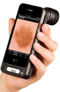
With new technologies, we believe that it is possible to diagnose skin cancer with mobile telephones. The department of Dermatology at Sahlgrenska University Hospital in Sweden is looking into these possibilities. With a HÜD attached to any smartphone, you can take a picture of a skin lesion you are worried about, then send it in with the First Derm iPhone app and get an answer from a dermatologist within 24 hours on whether it is suspicious or not .
Use our dermatology search engine to screen skin cancer
Learn about Dermoscopy
Colors & Structures in Melanocytic Lesions
Melanoma Specific Dermoscopic Structures


